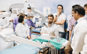
Endodontic microsurgery training course
Endodontic surgery is a discipline that is often poorly understood and under-practiced. Yet it is a more conservative technique than conventional endodontic removal, and an alternative to tooth extraction.
This course is designed to give dentists and endodontists an in-depth understanding of all the stages and techniques involved in endodontic surgery. These include indications and contraindications for endodontic microsurgery, anesthesia techniques, ergonomics, soft tissue management, incision layouts, osteotomy, apical resection, apical curettage, as well as post-operative follow-up and the use of bone grafts.

Indications and contraindications for endodontic microsurgery
Endodontic microsurgery may be indicated in the case of inadequate treatment: if retreatment is impossible, if obstacles exist (abutments, fractured instruments, perforation) and in the case of non-removable prostheses (long-span bridges or corono-radicular anchors).
It may also be indicated in the case of satisfactory treatment but persistent extra-root infection, or in the presence of a root cyst.
When canals are mineralized, or in the case of teeth with complex anatomies such as dens in dente, first-line treatment via the coronal route is technically complex and may prove to be disfiguring. Endodontic microsurgery is naturally indicated in these special situations.
Contraindications are of a general nature, linked to medical problems or local to the clinical situation. They are related to
- Anatomy (bone thickness, sinuses, chin foramen, palatal roots, inaccessibility).
- Periodontal status (mobility, crown/root ratio, marginal inflammation)
Steps and techniques covered in training
- Anesthesia in endodontic surgery: This section deals with pain management and patient comfort, covering the various techniques of local anesthesia. Anesthesia also ensures perfect hemostasis, which is essential in endodontic surgery.
- Ergonomics: Ergonomics is an often underestimated but essential aspect of endodontic surgery, influencing not only the quality of care but also the health of the practitioner. This section will explore ergonomic principles adapted to endodontic surgery, including the position of the patient, practitioner and assistant. The aim is to minimize fatigue and errors, optimizing working conditions for more precise and less stressful procedures.
- Soft tissue management and incision layouts: Soft tissue management is a crucial step in ensuring adequate access to the surgical area while preserving the integrity of surrounding structures. This section will cover various soft tissue manipulation techniques, including the use of retractors, bleeding management, and methods to minimize tissue trauma. The choice of incision route is also fundamental, as it influences visibility, access, healing and suture stability. We present different incision routes, their specific indications, and the aesthetic and functional considerations to be taken into account.
- Osteotomy, apical resection and apical curettage:
A 3 mm apical resection eliminates most of the lateral canals and exposes the isthmuses joining the main canals. The resection should be as perpendicular as possible to the long axis of the root. The resected surface must be observed at high magnification, using a micromirror to detect any micro-cracks. The main aim of root canal preparation is to remove the old filling material, eliminate infected dentine and create a space for a watertight filling. It is performed using ultrasonic inserts mounted on a piezoelectric generator.
This part of the course will examine apical resection techniques, the instruments used, and the precautions to be taken at each stage. Apical curettage will also be covered in detail. - Obturation: Two families of materials can be used for a retro obturation. Reinforced zinc oxide-eugenol cements, such as super EBA or IRM, and bioceramic materials, such as MTA, Total Fill or equivalent. Bioceramic materials can induce cement formation on contact, but no material is superior in terms of clinical success. The canal is filled with successive deposits, which are progressively condensed using microfillers. Microdrills of varying sizes up to 6 mm in length are available, enabling the material to be compacted deep into the root canal system. Fast-setting materials are polished at the end of the procedure.
- This section covers obturation techniques and criteria for choosing retrograde obturation material.
Post-operative follow-up: This section will cover drug prescriptions, pain management and infection prevention. Possible complications will also be discussed, with strategies for anticipating and managing them effectively. - Bone grafts: In some cases, endodontic surgery requires bone grafts to compensate for the loss of bone around the root after apical resection. This section will examine the indications for bone grafts, the types of material available (autografts, allografts, xenografts, synthetic biomaterials), and grafting techniques in endodontic surgery.
This training course on endodontic surgery will provide participants with an in-depth understanding of the various steps and techniques required to successfully carry out these complex procedures. From case selection and complication management to technological innovations and new surgical approaches, participants will leave with the skills and knowledge they need to improve their clinical practices and deliver high-quality care to their patients.
Training objectives
The following are the training objectives, formulated with action verbs:
- Understand the indications and contraindications of endodontic microsurgery to determine the appropriate clinical cases for this procedure.
- Master anesthesia techniques for endodontic surgery, selecting appropriate anesthetic agents and applying optimal administration methods.
- Apply ergonomic principles to improve surgical quality, reduce fatigue and minimize clinical errors.
- Perform incision layouts and soft-tissue management to ensure optimal access to the surgical area while preserving surrounding structures.
- Perform the various technical stages of osteotomy, apical resection and apical curettage, using specific instruments and following protocols to minimize trauma and promote healing.
- Prepare and perform the necessary post-operative care to ensure rapid recovery and prevent post-surgical complications.
- Use optical aids, such as the operating microscope, to effectively identify and treat anatomical and pathological features during surgery.
- Perform bone grafts when necessary, selecting appropriate materials and applying techniques to maintain the structural and functional integrity of the surgical site.
- These objectives are designed to ensure that participants acquire a comprehensive understanding and practical competence in endodontic microsurgery.